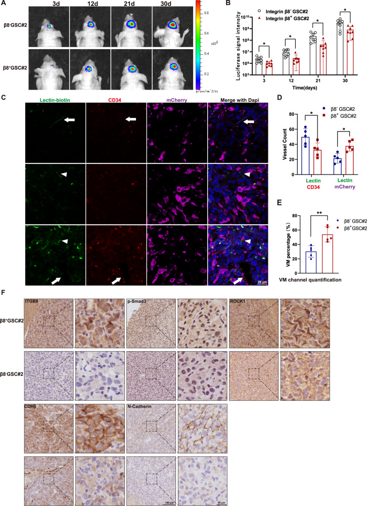
Fig. 6. β8 integrin contributes to VM formation in intracranial GBM xenograft.
A Luminescent imaging of representative nude mice xenografts from mCherry-labeled β8+ (n = 8) or β8− GSC#2 (n = 8) at day 3, 12, 21 and 30. B Luminescent signal intensity of GBM-bearing mice in two groups were evaluated. C Representative immunofluorescence images of vascular channels lined by ECs or tumor cells. Arrows indicate the regular CD34+ vessels, while arrowheads indicate CD34-/Lectin+ vessels. Scale bar = 20 μm. D Quantification of lectin (+) vessels stained positively by CD34 or mCherry. E Percentage of VM vessels in β8+ or β8− integrin xenografts. F IHC staining of ITGB8, p-Smad3, ROCK1, CDH5 and N-Cadherin in β8+ and β8− integrin xenografts. Results are represented as mean ± SD of biologically triplicate assays. *p < 0.05, **p < 0.01, ***p < 0.001.

