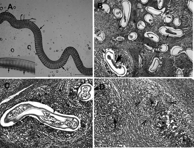
Fig. 1.
a A fragment of the worm dissected out of the nodule showing marked and straight transverse ridges on the outer surface of the cuticle with small protuberances on the both lateral sides (arrow), Bar = 100 μm. Inset picture, showing the ridges (arrow), Bar = 20 μm. b Histological section of the subcutaneous nodule showing multiple adult female worms of O. flexuosa (arrows) with few males, the parasites are surrounded by homogenous eosinophilic material; inset picture showing the engorged uterus with the parasite, H&E, Bar = 200 μm. c Onchocerca females (arrow) with numerous microfilariae in the uterus (arrow heads) and granulomatous reaction surrounding the parasite (star) by H&E staining. Bar = 100 μm. d Degenerated filarial parasites represented by homogenous eosinophilic material (star) and severe granulomatous reaction (arrow) of macrophages and lymphocytes and eosinophilia (arrowheads) by H&E staining. Bar = 100 μm

