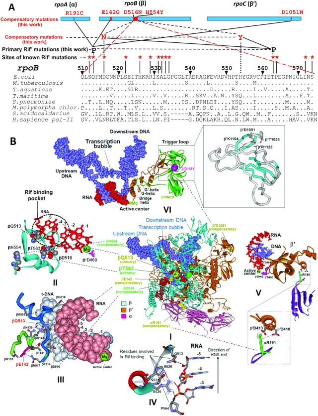
Figure 1.
Structural and genetic context of rifampicin resistance determining region (RRDR) of bacterial RNA polymerase and location of the rifampicin resistant and compensatory mutations selected in this study. (A) Top, location of the selected mutations in RNAP subunits. Bottom, sequence alignment of RRDR from various organisms. Positions of known Rifr mutations along with those identified in this work (black font) and corresponding compensatory substitutions (red font) are indicated. (B) Locations of the selected mutations in RNAP TEC (panel I). Panels II-VI represent enlarged images of I showing the location of particular mutations. Primary Rifr mutations and corresponding compensatory mutations have the same color coding. Green sphere represents catalytic Mg2+ ion of the active center. Structure of E. coli RNAP TEC is from Supporting Information of (54).

