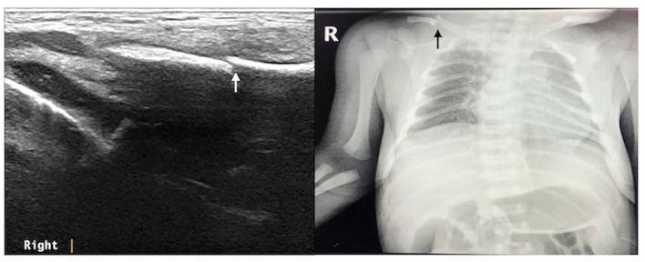
Figure 2.
Right clavicle fracture. The infant was G2P1 with a gestational age of 39+1 weeks, vaginal delivery, and birth weight of 4,140 g. The patient was admitted to the hospital 3 h after birth due to intrauterine pneumonia. Physical examination showed that the bilateral clavicles were asymmetrical and that the left side was smooth, while the right side had obvious bone rubbing. Ultrasound revealed interrupted continuity of the cortical bone of the right clavicle and broken end formation and dislocation. The fracture of the clavicle was confirmed by chest X-ray examination.

