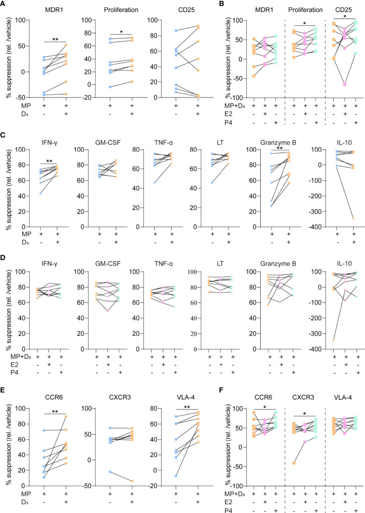
Figure 2.
The suppressive capacity of steroid hormone cocktails on glucocorticoid-resistant Th17.1 cells. (A) MDR1 expression, proliferation rates (CFSE-) and CD25 surface expression (MFI) by healthy donor anti-CD3/CD28-stimulated Th17.1 cells exposed to MP with and without D3 as determined by flow cytometry (n = 8). Percentages are relative to their appropriate vehicle control. (B) The same parameters as in A) for healthy donor anti-CD3/CD28-stimulated Th17.1 cells exposed to MP+D3 with and without E2 or P4 as determined by flow cytometry (n = 8). Percentages are relative to their appropriate vehicle control. (C) Amount (pg/ml) of IFN-γ, GM-CSF, TNF-α, LT, granzyme B, and IL-10 measured in the supernatants of healthy donor anti-CD3/CD28-stimulated Th17.1 cells exposed to MP with and without D3 as determined by Luminex (n = 8 per group). (D) Amount (pg/ml) of IFN-γ, GM-CSF, TNF-α, LT, granzyme B, and IL-10 measured in the supernatants of healthy donor anti-CD3/CD28-stimulated Th17.1 cells exposed to MP+D3 with and without E2 or P4 as determined by Luminex (n = 8 per group). (E) CCR6, CXCR3 and VLA-4 surface expression on healthy donor anti-CD3/CD28-stimulated Th17.1 cells exposed to MP with and without D3 as determined by flow cytometry (n = 8). Percentages are relative to their appropriate vehicle control. (F) The same parameters (as in E) for healthy donor anti-CD3/CD28-stimulated Th17.1 cells exposed to MP+D3 with and without E2 or P4 as determined by flow cytometry (n = 8). Percentages are relative to their appropriate vehicle control. Data were compared using either Wilcoxon rank-sum or (D) Friedman tests with the false discovery rate of Benjamini, Krieger and Yekutieli correction. *p < 0.05 and **p < 0.01. “D3 = 1,25(OH)2D3”, “E2, Estradiol”; “MFI, median fluorescence intensity”; “MP, methylprednisolone” and “P4, progesterone”.

