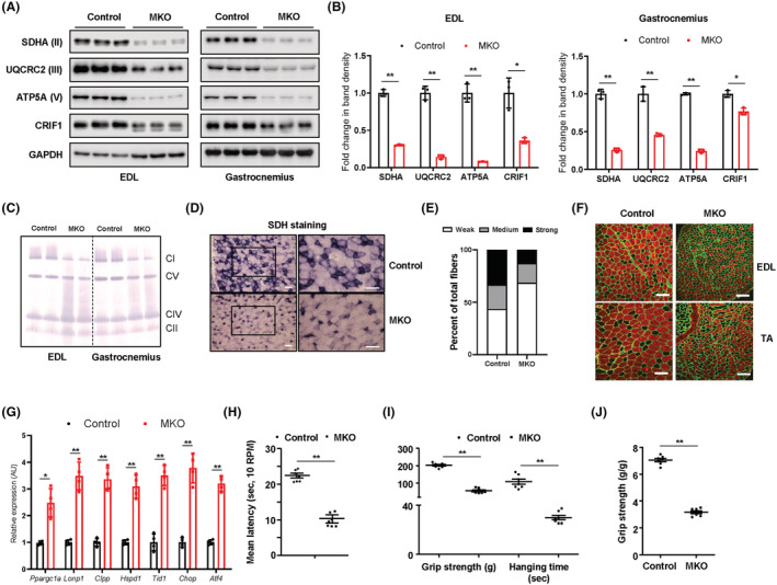
Figure 1.

MKO mice show impairment in OxPhos and deterioration in physical performance. (A,B) Representative western blots and band density measurements for OxPhos complex subunits and CRIF1 in the EDL and gastrocnemius of chow‐fed control and MKO mice at 14 weeks of age; n = 3. (C) Representative blots showing BN‐PAGE of the assembled OxPhos complex in EDL and gastrocnemius from chow‐fed control and MKO mice at 14 weeks of age. (D) Transverse EDL sections were stained histochemically for SDH to identify oxidative muscle fibres at 14 weeks of age. Scale bar, 100 μm. (E) Quantification of unstained fibres in the EDL of controls and MKO mice at 14 weeks of age; n = 5. (F) Cross‐sections of the EDL and TA muscle from 10‐week‐old control and MKO mice were subjected to immunohistochemical staining for MyHC2b (red) and laminin (green). Scale bar, 100 μm. (G) Relative expression of mRNA encoding genes related to mitochondrial stress response from EDL in 14‐week‐old control and MKO mice; n = 4. (H) Latency to fall in rotarod test at 0.00336 g; n = 7. (I) Forelimb grip strength and time to fall in the wire hanging assay for control and MKO mice; n = 10. (J) Grip strength normalized to the body weight of control and MKO mice; n = 10. Data are expressed as the mean ± standard error of the mean. Statistical significance was analysed by unpaired t‐tests. *, P < 0.05 and **, P < 0.01 compared with the indicated group.
