
FIGURE 2.
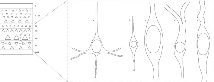
Laminar location and morphological characteristics of human VENs. The diagram on the left shows the location of VENs in the deep part of layer V. The images on the right show the morphology of a pyramidal neuron (a), a fusiform neuron in layer VI (b) and three morphological variants of VENs (c, d and e). The size of the soma of these illustrated neurons maintains the relative proportions found in cortical tissue