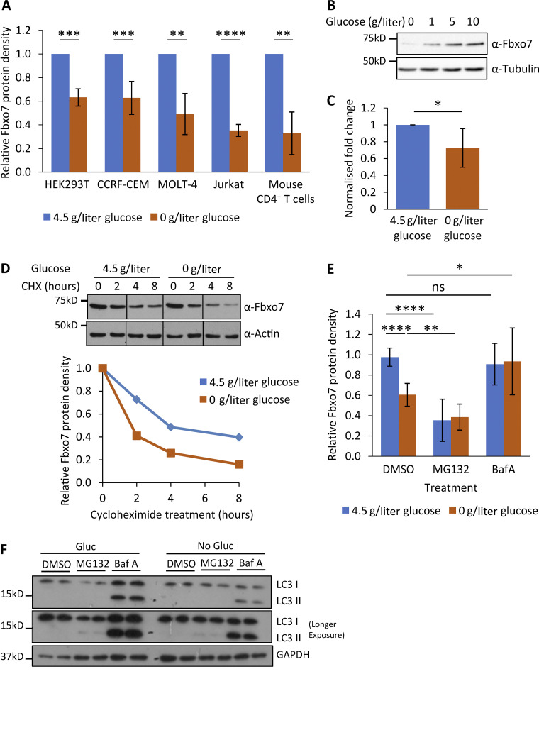
Figure 5.
Regulation of Fbxo7 by glucose availability. (A) Relative Fbxo7 protein following 48 h of glucose starvation, analyzed by immunoblot and quantified (n ≥ 3). (B) CCRF-CEM cells cultured for 48 h with a titration of glucose, then lysed and analyzed by immunoblot (n = 3). (C) Relative Fbxo7 mRNA in naive murine CD4+ T cells, incubated for 4 h in 0 or 4.5 g/liter glucose (n = 6). (D) CHX chase in CCRF-CEM cells following the removal (0 g/liter) or maintenance (4.5 g/liter) of glucose. Fbxo7 protein measured by immunoblot (top) and quantified (bottom; representative of n = 3). (E) CCRF-CEM cells were treated with DMSO, 10 μM MG132, or 200 nM BafA1 (BafA) immediately following glucose removal (0 g/liter) or maintenance (4.5 g/liter). Fbxo7 protein measured by immunoblot and quantified (n = 7). (F) Image of representative immunoblot for LC3I/II from samples from E (n = 4). *, P < 0.05; **, P < 0.01; ***, P < 0.001; ****, P < 0.0001. Source data are available for this figure: SourceData F5.

