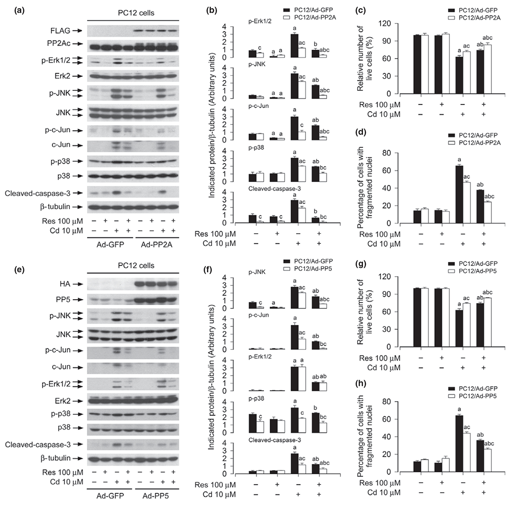
Fig. 7.

Over-expression of PP2A and PP5 strengthens resveratrol inhibition of Cd-induced activation of extracellular signal-regulated kinases 1/2 (Erk1/2), JNK and/or p38, as well as cell death. PC12 cells, infected with Ad-PP2A, Ad-PP5, or Ad-GFP (as control), were pre-treated with/without resveratrol (Res, 100 μM for 1 h, and then exposed to Cd (10 μM for 4 h (for western blotting) or 24 h (for live cell analysis, 4′,6-diamidino-2-phenylindole, DAPI staining). (a and e) Cell lysates were subjected to western blot analysis using indicated antibodies. The blots were probed for β-tubulin as a loading control. Similar results were observed in at least three independent experiments (a and e), and blots for p-JNK, p-c-Jun, p-Erk1/2, p-p38, cleaved-caspase-3 were semi-quantified (b and f). (c and g) Live cells were detected by counting viable cells using trypan blue exclusion. (d and h) The percentages of apoptotic cells with fragmented nuclei were quantified by DAPI staining. Results are presented as mean ± SE, n = 5. Using one-way anova or Student’s t-test, ap < 0.05, difference with control group; bp < 0.05, difference with 10 μM Cd group; cp < 0.05, Ad-PP2A group or Ad-PP5 group versus Ad-GFP group.
