
FIGURE 1.
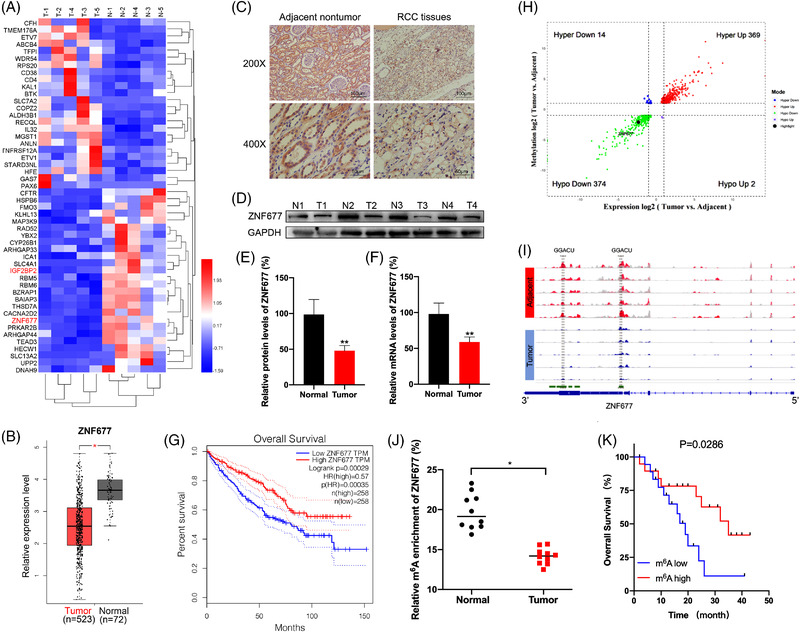
Downregulation of ZNF677 is associated with unfavourable prognosis and decreased m6A methylation modification levels in renal cell carcinoma (RCC). (A) Heatmap of differentially expressed genes in five pairs of matched RCC tissues and adjacent normal tissues by RNA‐seq. (B) Boxplot showing ZNF677 mRNA expression in 523 RCC tumour tissues (red plot) and 72 normal tissues (grey plot) (http://gepia2.cancer‐pku.cn). (C) Immunohistochemical analysis of ZNF677 performed on RCC tissues and adjacent normal tissues. (D) ZNF677 expression was determined by Western blot analysis in RCC tissues and their matched noncancerous tissues. GAPDH was used as loading control. (E) The quantitative illustration of the levels of ZNF677 protein in (D) was used for densitometry to measure the density of the corresponding bands on the Western blot analysis. (F) qRT‐PCR assay verified the expression of ZNF677 in matched RCC tissues and adjacent normal tissues. (G) Kaplan–Meier survival plot of RCC patients (n = 516) stratified by low (blue line) and high (red line) ZNF677 expression. (H) Four quadrant diagrams show the differentially methylated genes and differentially expressed genes in five pairs of matched RCC tissues and adjacent normal tissues detected by MeRIP‐seq and RNA‐seq. (I) Integrative Genome Viewer (IGV) software showed the m6A peaks within ZNF677 mRNA in five pairs of matched RCC tissues and adjacent normal tissues. (J) m6A enrichment on ZNF677 mRNA in 10 pairs of matched RCC tissues and adjacent normal tissues detected by MeRIP‐qPCR. (K) Kaplan–Meier survival analyses of the relationship between the levels of m6A of ZNF677 and survival time in RCC patients. *p < .05 or **p < .01 indicates a significant difference between the indicated groups