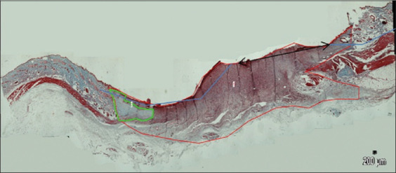
Figure-5.

Wound histology on day 21 (Masson’s trichrome staining, 40×, pictures were combined using software PTGui (Pro software by New House Internet Service B.V; Rotterdam, the Netherlands); red line shows the wound area; green line shows collagen area; two-headed arrow shows the edges of re-epithelization area.
