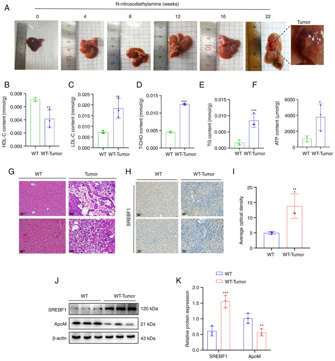
Figure 1.
High expression of SREBF1 in N-nitrosodiethylamine-induced mouse hepatocellular carcinoma tissues. (A) Images of livers from mouse models with N-nitrosodiethylamine-induced liver cancer tumors. (B-F) A test kit was utilized to detect the content of (B) HDL-C, (C) LDL-C, (D) T-CHO, (E) TG and (F) ATP in liver cancer tissue of WT mice and liver tissue from healthy mice. (G) H&E staining revealed the morphology of liver tissue in mice prior to and after induction with N-nitrosodiethylamine (scale bar, 100 µm). (H) SREBF1 expression levels were determined using immunohistochemistry (scale bar, 50 µm). (J and K) Western blot analysis was performed to evaluate the expression levels of SREBF1 and ApoM in liver cancer tissue of WT mice and liver tissue from healthy mice. (J) Representative western blots and (K) quantified results. Analysis in each group was performed three times in parallel. *P<0.05, **P<0.03, ***P<0.01 vs. WT. HDL-C, high-density lipoprotein cholesterol; LDL-C, low-density lipoprotein cholesterol; T-CHO, total cholesterol; TG, triglyceride; WT, wildtype; SREBF1, sterol regulatory element-binding protein 1; ApoM, apolipoprotein M.

