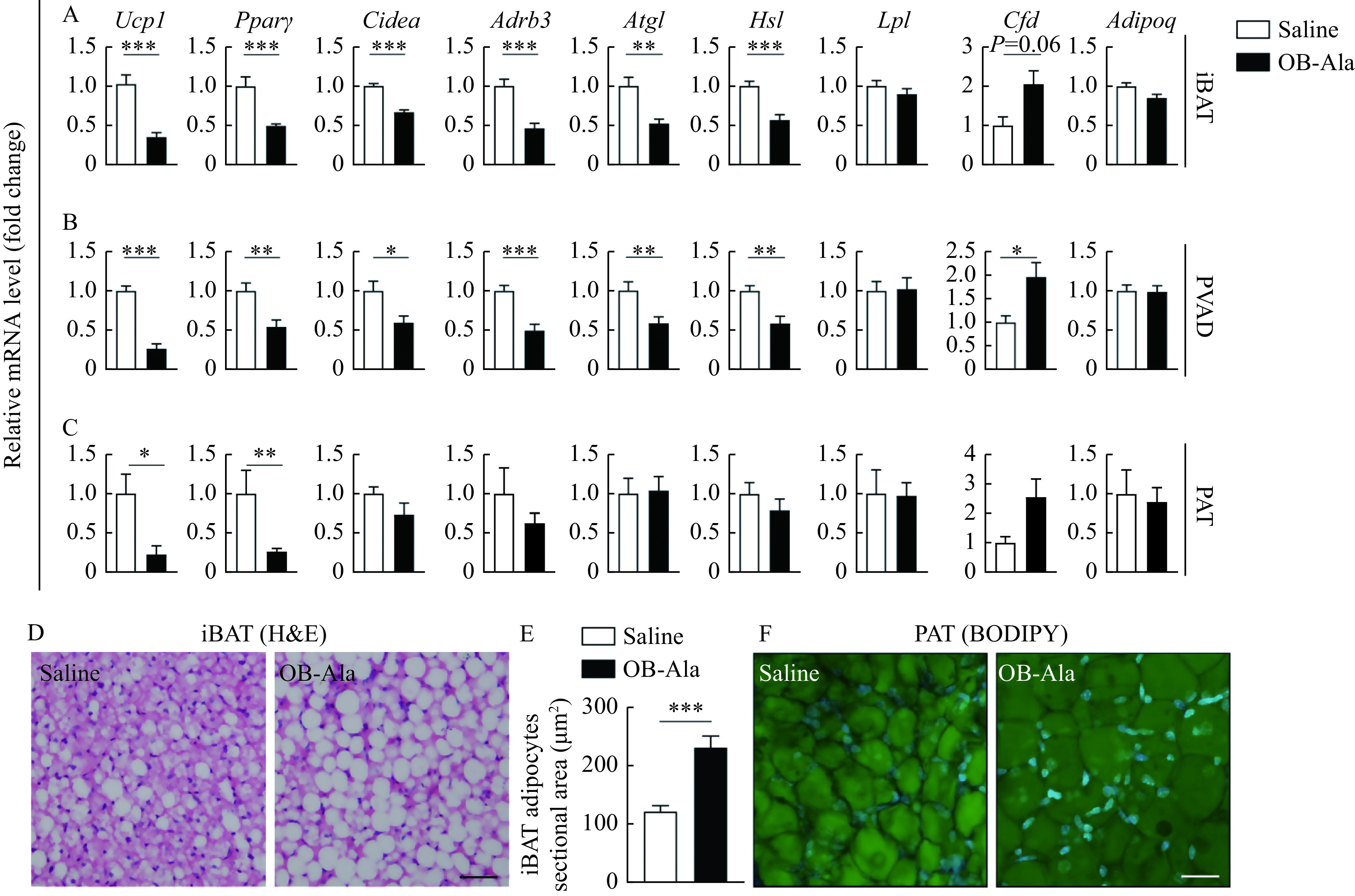
Figure 2.

OB-Ala inhibited thermogenesis of brown adipose tissue.
Mice received daily i.p. injection of OX2R agonist OB-Ala (16 nmol/kg) or saline for 3 weeks. Brown adipose tissues were analyzed after 3 weeks of treatment. A–C: Relative gene expression of Ucp1, Pparγ, Cidea, Adrb3, Atgl, Hsl, Lpl, Cfd, and Adipoq in iBAT (A), PVAD (B), and PAT (C) of mice. Rps18 was used as the internal reference. D: Representative images of H&E staining of iBAT. E: Sectional area of iBAT adipocytes. F: Representative images of BODIPY staining of PAT.*P<0.05,**P<0.01,***P<0.001.n=7 for saline; n=9 for OB-Ala. Scale bar = 25 μm (D and F). Data arepresented as mean±SEM. Statistical significance was determined by unpairedt-test. OX2R: orexin receptor type 2; OB-Ala: [Ala11, D-Leu15]-OxB; iBAT: intrascapular brown adipose tissue; PVAD: perivascular adipose tissue; PAT: pericardial adipose tissue; BODIPY: borondipyrromethene; H&E: hematoxylin and eosin.
