
Figure 4.
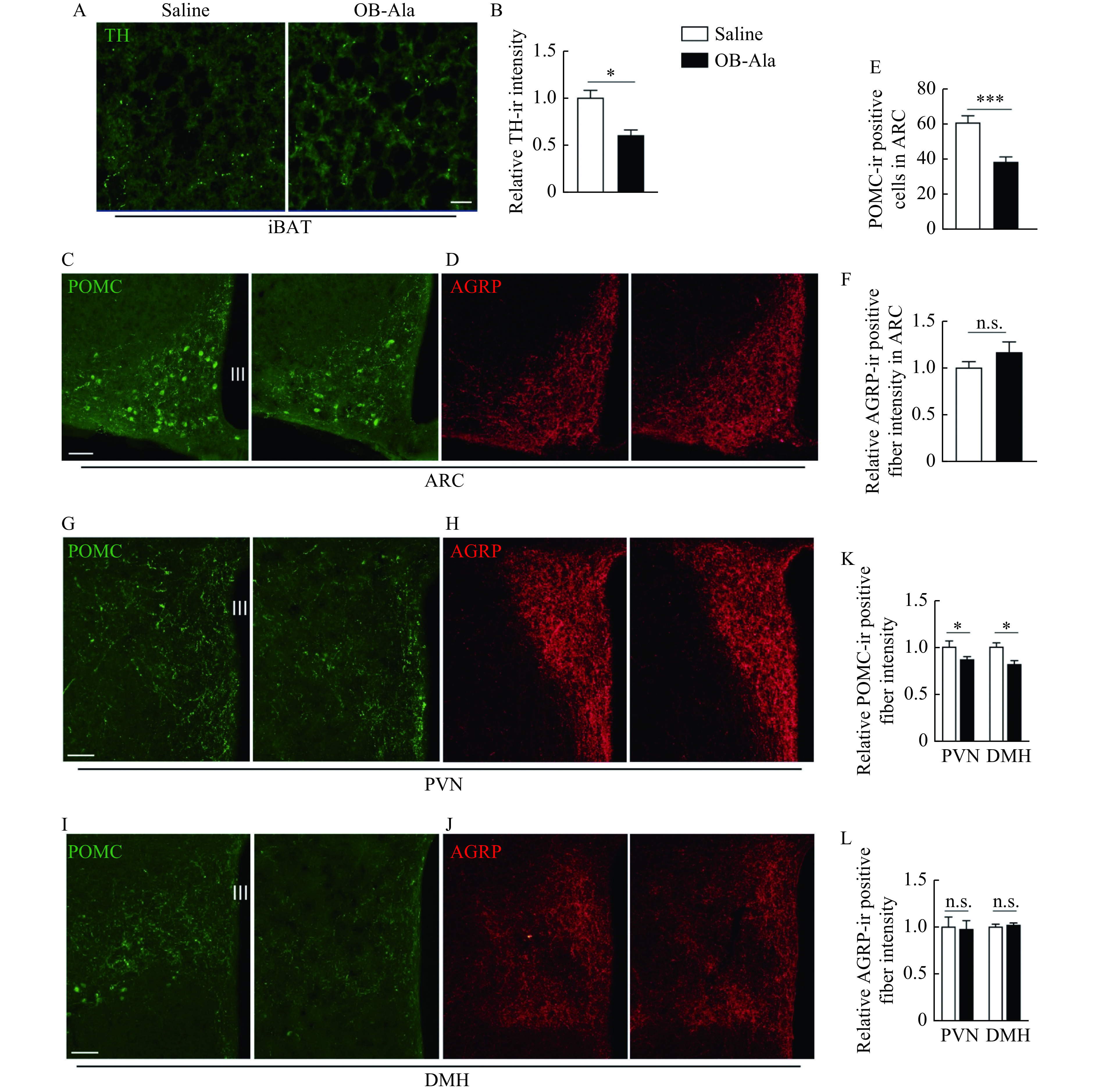
OB-Ala reduced sympathetic innervation of iBAT and POMC expression in the hypothalamus.
Mice received daily i.p. injection of OX2R agonist OB-Ala (16 nmol/kg) or saline for 3 weeks. A: TH fibers distribution in iBAT from mice after 3 weeks of treatment. B: Quantification of TH-immunoreactivity (TH-ir) positive fiber in panel A. C and D: POMC (C) and AGRP staining (D) in ARC region from mice after 3 weeks of treatment. E: Quantification of POMC-immunoreactivity (POMC-ir) positive cells in panel C. F: Quantification of ACRP-immunoreactivity (ACRP-ir) positive fibers in panel D. G and H: POMC (G) and AGRP (H) staining in PVN. I and J: POMC (I) and AGRP (J) staining in DMH. K: Quantification of panels G and H. L: Quantification of panels I and J. *P<0.05,***P<0.001.n=4 for saline; n=6 for OB-Ala. Data arepresented as mean±SEM. Statistical significance was determined by unpaired t-test. Scale bars: 20 μm (A) and 40 μm (C, G, and I). OX2R: orexin receptor type 2; iBAT: intrascapular brown adipose tissue; TH: tyrosine hydroxylase; POMC: proopiomelanocortin; AGRP: agouti-related peptide; ARC: arcuate nucleus; PVN: paraventricular nucleus; DMH: dorsal medial hypothalamus; III: third ventricle.