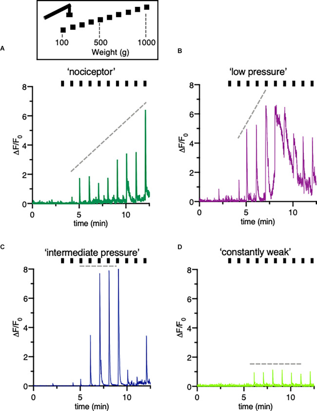
Figure 4.
Classification of different type of DRG neurons responsive to increasing pressure. (A–D) Schematic drawing of the mechanical stimulation protocol and example trace indicating a variety of fluorescence responses taken from individual neurons of a mouse treated with vehicle only. The hindpaw was stimulated with increasing pressure started at 100 g to a maximum of 1,000 g. Different patterns of changes in intracellular calcium concentrations reflect distinctive cell types, here called “nociceptors” (A) “low pressure” responders (B) “intermediate pressure” responders (C) and “constant weak” (D) responders. Ticks correspond to mechanical stimulation protocol.

