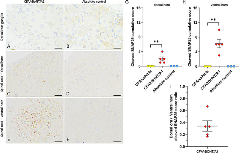
Figure 8.
Cleaved SNAP25 in the dorsal root ganglion (DRG) and the lumbar spinal cord. (A,B) Representative images of the dorsal root ganglion (DRG) and (C–F) of the spinal cord of mice treated with CFA/rBoNT/A1 or in untreated controls. Cleaved SNAP25 was absent in the DRG (despite higher background staining), while being present in the nerve ending-like structures in the ipsilateral dorsal (C) and ventral (E) horns of the lumbar spinal cord of CFA/rBoNT/A1-treated animals, but not in absolute controls. Bars (A,B) (20 μm), (C–F) (50 μm). (G–I) Quantification of cleaved SNAP25 in the lumbar spinal cord in CFA/rBoNT/A1-treated animals compared to CFA/vehicle and absolute controls. c-SNAP25 staining was moderate in the dorsal horn (G) and high in the ventral horn of the spinal cord (H) while being absent in CFA/vehicle-treated animals and in absolute controls. (I) The dorsal horn/ventral horn ratio of c-SNAP25 (< 1) indicates higher targeting of ventral vs. dorsal horn by rBoNT/A1. N = 5/group (CFA/rBoNT/A1, CFA/vehicle) and N = 3 (absolute control). Mean ± SEM. **p < 0.01 CFA/rBoNT/A1 vs. CFA/vehicle; Mann-Whitney test.

