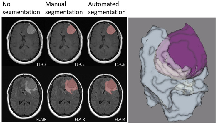
Figure 1.
Manual and automated segmentation comparison in a meningioma of the left frontal convexity. The sharply demarcated lesion demonstrates intense contrast enhancement. Vasogenic edema of the surrounding white matter is also evident. Manual and automated segmentation are correctly matched. Three-dimensional rendering of the segmented tumor and edema volumes is also presented. T1-CE: contrast-enhanced T1-weighted imaging; FLAIR: fluid attenuated inversion recovery. Adapted from Ref. [13], under the terms of the Creative Commons Attribution-NonCommercial 4.0 License.

