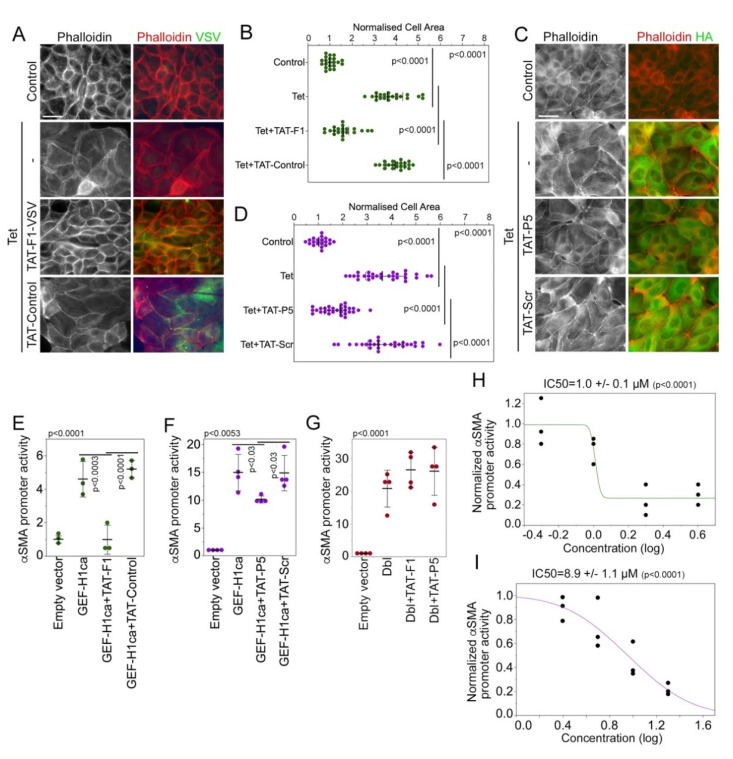
Figure 3.
Membrane-permeable GEF-H1 inhibitors impede overexpressed GEF-H1. (A–D) TAT-F1 or TAT-Control (A,B: 4 µM), or TAT-P5 or TAT-Scr (C,D: 20 µM) were incubated overnight with MDCK cells induced to overexpress HA-GEF-H1. The cells were then analyzed by staining for F-actin and the VSV-epitope to detect cells expressing the VSV-tagged CTD fragments (A) or F-actin and the HA-epitope to monitor HA-tagged GEF-H1 expression (C). Cell areas were measured to assess cell spreading (B,D). The quantifications show values for cell sizes measured over 2 (B) or 3 (D) experiments. p-values were calculated with Wilcoxon and Mann-Whitney tests. (E–I) HEK 293 cells were co-transfected with a plasmid containing an αSMA promoter driving firefly luciferase expression, one with a CMV promoter stimulating renilla luciferase expression, and a plasmid encoding GEF-H1-CA. The cells were incubated with TAT-F1 (E,G 4 µM) or TAT-P5 (F,G 20 µM) or with the concentrations indicated (H, TAT-F1; I, TAT-P5) overnight. The activity of the luciferases was then measured and the activity of the αSMA promoter was expressed as a ratio between firefly and renilla luciferase activity. Shown are independent determinations, and p-values derived from ANOVA and two-sided t-tests. Scale bars, 20 μm.

