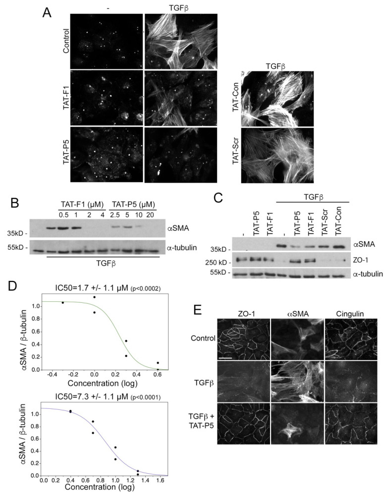
Figure 4.
GEF-H1 antagonists inhibit epithelial mesenchymal transition induced by TGFβ in primary retinal pigment epithelial (RPE) cells. (A–D) Primary RPE cells were incubated without or with TGFβ for five days in the presence of TAT-F1, TAT-P5, TAT-scr, or TAT-control expression of αSMA positive cells was analyzed by immunofluorescence (A) or immunoblotting (B–D; α-tubulin was used as a loading control). The graphs in panel D show quantifications of concentration-dependent expression of αSMA normalized by α-tubulin expression derived from densitometric scanning of the immunoblots. Note, that GEF-H1 inhibitors strongly reduce TGFβ-induced αSMA expression in a concentration-dependent manner (TAT-F1, upper and TAT-P5, lower graph). Panel C also shows immunoblots for ZO-1, revealing attenuation of ZO-1 downregulation by GEF-H1 inhibitors. (E) Expression of the junctional markers ZO-1 and cingulin, as well as αSMA was analyzed by immunofluorescence after incubating without inhibitors or with either TAT-P5. Note, TAT-P5 inhibits TGFβ induced αSMA expression and loss of junctional ZO-1 and cingulin staining. Scale bar, 30 μm.

