Figure 5.
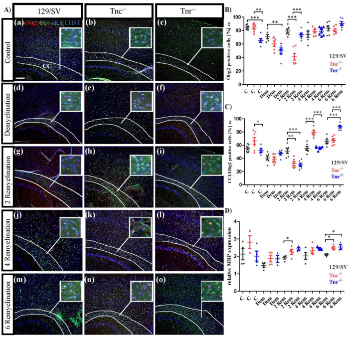
Differential maturation of oligodendrocytes in Tnc−/− and Tnr−/− genotypes after cuprizone treatment. (A(a–o)) Sagittal sections of 129/SV, Tnc−/− and Tnr−/− mice treated with cuprizone were immunohistochemically stained with antibodies against CC1 (green) and Olig2 (red) in the five different conditions, control (C), demyelination (DM), 2-, 4- and 6 weeks of remyelination (RM2, RM4, and RM6). Hoechst was used as a marker for cell nuclei (blue). Images show the caudal part of the corpus callosum (CC) above the hippocampus (HC), circle triangle squares show a better visualization of the cells. Fewer oligodendrocytes were detected in Tnr−/− condition in comparison to the 129/SV wildtype and the Tnc−/− genotype (A(a–c),B). CC1 staining was reduced by cuprizone treatment and recovered after 2, 4 and 6 weeks of remyelination (A(d–o)). In the early stage of demyelination after 2 weeks, lowest number of oligodendrocytes was detected in Tnc−/− mice, and the highest number of immature oligodendrocytes was detected in 129/SV wildtype mice (A(g–i),C). RT-PCR analysis of MBP expression revealed a higher myelin expression in both knockouts at the earliest (2 weeks) and latest (6 weeks) stages of recovery, reflecting a more extensive remyelination in Tnc−/− and Tnr−/− mice (D). Data are presented as mean ± SEM and statistical significance (p ≤ 0.05 *, p ≤ 0.01 **, p ≤ 0.001 ***) was assessed using the ANOVA and Tukey’s multiple comparison test (control, demyelinated, remyelinated). Four animals were used for each group and genotype (N = 4).

