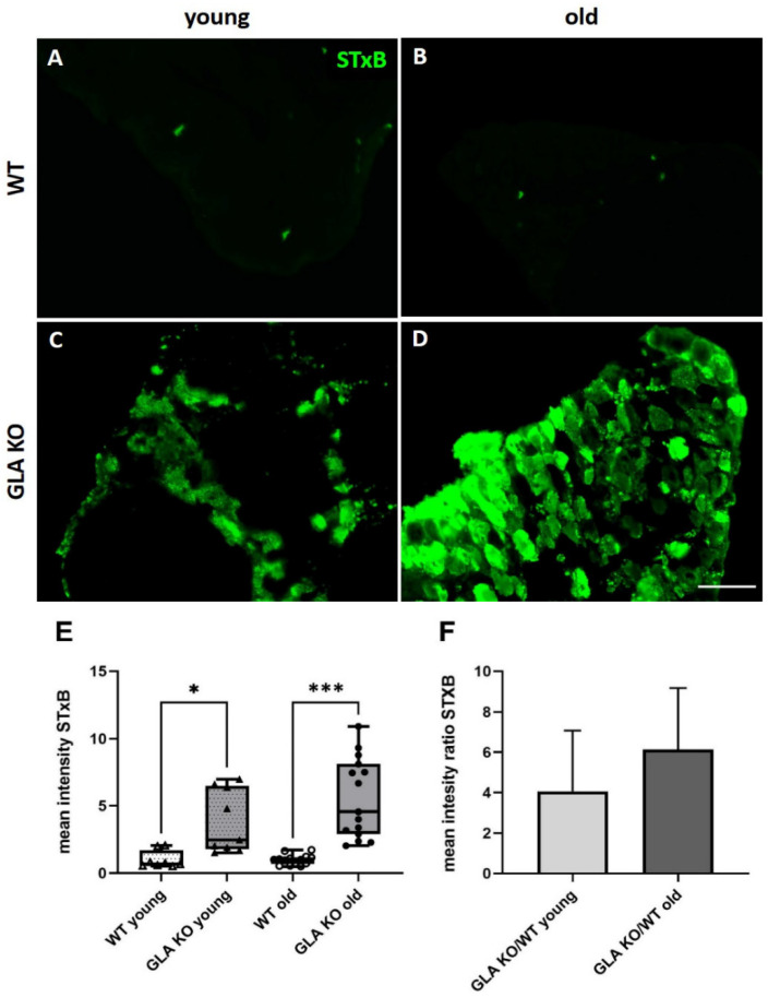
Figure 1.
Mean intensity of STxB::555 in murine DRG. (A–D) Representative photomicrographs of Gb3 accumulations in DRG of young WT (A), old WT (B), young GLA KO (C), and old GLA KO mice (D) visualized with STxB::555 (green). (E) Intensity measurements of STxB::555 signal in murine DRG of young WT (△, n = 8), young GLA KO (▲, n = 9), old WT (◯, n = 14), and old GLA KO mice (●, n = 15). (F) Mean intensity GLA KO/WT ratio of STxB::555 signal for young and old murine DRG. Abbreviations: DRG = dorsal root ganglion; GLA KO = alpha-galactosidase A knockout; STxB = Shiga toxin 1, subunit B; WT = wildtype. Scale bar: 100 µm. * p < 0.05, *** p < 0.001.

