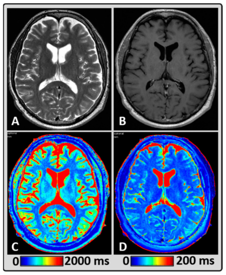
Figure 1.
Representative MRI data from a 58-year-old male BM patient treated with focal radiation therapy (RT), showing normal-appearing brain tissue. (A,B) standard T2w and post-contrast T1w images depicting the anatomical structures, (C,D) T1 and T2 maps were estimated using MAGnetic resonance image Compilation (MAGiC) for a single slice exhibiting gray matter (GM), white matter (WM), and cerebrospinal fluid (CSF).

