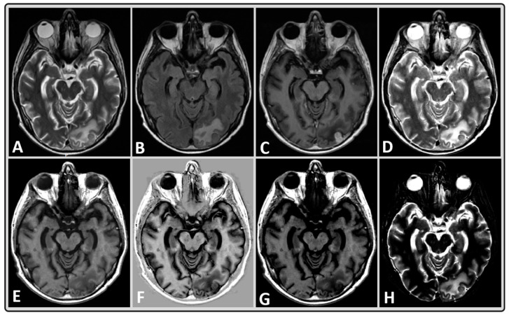
Figure 2.
Representative MRI data of a 40-year-old male BM patient treated with whole-brain RT using standard and synthetically-generated (MAGiC) images. (A–C) T2w, T2w fluid attenuated inversion recovery (FLAIR) and T1w post-contrast images, respectively, from standard clinical imaging. (D–H) T2w, T1w, phase-sensitive inversion recovery (PSIR), T1w FLAIR, and short tau inversion recovery (STIR) images, respectively, from MAGiC.

