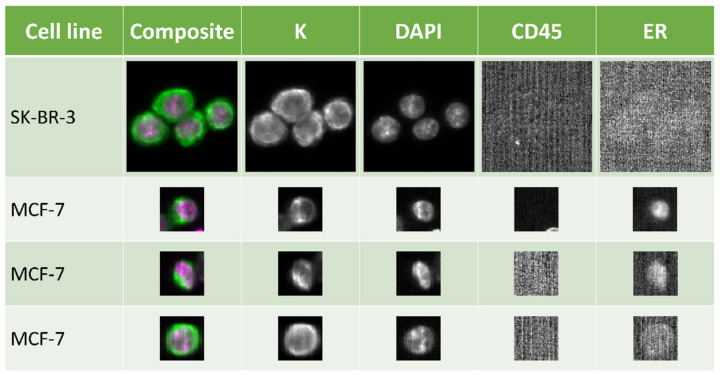
Figure 1.
ER expression detected in breast cancer cells by the CellSearch System. SK-BR-3 (top row) is an ER-negative cell line and was used as a negative control. MCF-7 is ER-positive and examples are given for strongly positive, (second row), weakly positive, (third row), and (bottom row) nuclear staining. Standard selection markers of the CellSearch System for tumor cells are keratin (K) and DAPI, whereas CD45 is used as an exclusion marker. Automatic low signal compensation leads to high background signal seen in CD45 and ER-negative scans. Pictures were taken using 10× magnification.

