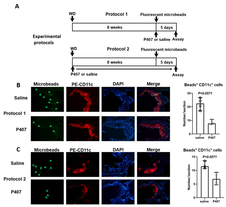
Figure 6.
CD11c+ (CD36+) monocyte infiltration into atherosclerotic plaques. (A) Experimental protocols for WD feeding, P407 or saline injection, and injection of fluorescent microbeads (to label CD11c+ monocytes) in Ldlr−/− mice. Representative images of aortic root showing infiltration of microbead (green)-labeled CD11c+ monocytes into atherosclerotic plaques and staining for CD11c (red), DAPI (blue), and quantitation of labeled CD11c+ monocyte infiltration into plaques in protocols (B) 1 and (C) 2. n = 3–4 mice/group. Data are shown as median with 95% confidence interval and were analyzed by the Mann–Whitney test.

