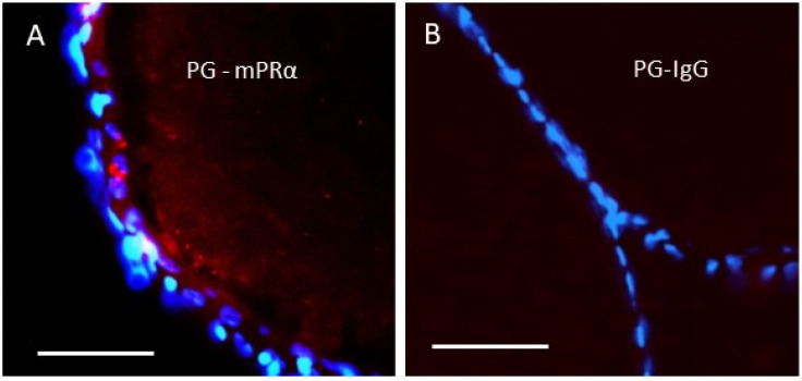
Figure 2.
In situ proximity ligation analysis (PLA) of the association of mPRα with a PGRMC1 in zebrafish oocytes using zebrafish mPRα and PGRMC1 (PG) antibodies. (A) Close proximity of mPRα and PGRMC1 (<40 nm) in the image are shown as red dots. (B) PLA using the PGRMC1 antibody and IgG as a negative control showing the absence of red dots in the image. The nuclei (blue) of the follicle cells surrounding the oocyte are stained with DAPI. Scale bar 100 µm. Produced from Aizen et al. [34] with permission.

