
Fig. 1. Design and characterization of PPNs.
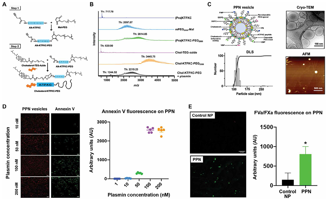
(A) Shown is the two-step process for synthesis of cholesterol-KTFKC-PEG using alkyne-terminated plasmin-cleavable peptide (Alk-KTFKC), maleimide-terminated polyethylene glycol (Mal-PEG), and azide-terminated cholesterol-triethylene glycol (cholesterol-TEG-azide). Cholesterol-KTFKC-PEG was incorporated into PPNs providing a PEG cloak that could be cleaved by plasmin. (B) Matrix-assisted laser desorption ionization–time-of-flight mass spectrometry was used to characterize synthesized cholesterol-KTFKC-PEG and its plasmin-induced degradation (Th, theoretical mass). (C) Shown is the design of PPN vesicles and their size characterization by dynamic light scattering (DLS), cryo–transmission electron microscopy (cryo-TEM), and atomic force microscopy (AFM). The PPN diameter ranged from 100 to 150 nm. (D) Shown are representative images and fluorescence intensity quantification of fluorescently labeled annexin V that was bound to exposed phosphatidylserine at the surface of immobilized PPNs after exposure to plasmin. (E) Shown are representative images and fluorescence intensity quantification of Alexa Fluor 488–labeled antibody bound to factors FVa and FXa (green fluorescence) of the prothrombinase complex that was assembled at the surface of PPNs with exposed phosphatidylserine but not on control nanoparticles (Control NP) without exposed phosphatidylserine. *P ≤ 0.05, two tailed t test.