
Fig. 3. PPNs enhance human plasma and whole blood clot stability under fibrinolytic conditions.
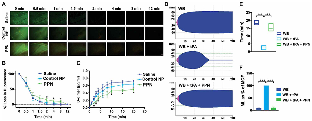
(A to C) Shown is microfluidic analysis of human plasma clots exposed to tissue plasminogen activator (tPA) to create a fibrinolytic environment. Addition of PPNs, with exposed phosphatidylserine in response to plasmin (PPN) in human plasma, delayed clot lysis as indicated by fibrin green fluorescence (A and B) and reduced D-dimers in the clot lysate (C) compared to control nanoparticles (Control NP). (D to F) Shown is rotational thromboelastometry analysis of human whole blood in the presence of tPA. Addition of PPNs, with exposed phosphatidylserine in response to plasmin (PPN), enhanced clot stability as demonstrated by the MCF maintenance time (E) and reduced maximum lysis (ML) (ML as % of MCF) (F). *P ≤ 0.05, **P ≤ 0.01, and ***P ≤ 0.001, two-way ANOVA with Tukey’s multiple comparisons for microfluidic data and one-way ANOVA with Tukey’s multiple comparisons for rotational thromboelastometry data.