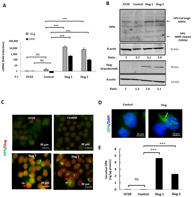
Figure 3.
Slug/SNAI2 promotes the expression and secretion of OPN. HT-29 cells were stably transfected with a plasmid coding for Slug or with an empty expression vector (control). (A) The expression of Slug and OPN mRNA was determined by qRT-PCR, and the results were normalized to HT-29. (B) The expression of Slug and osteopontin protein was determined by Western blot analysis with β-actin as loading control. (C) Cells were stained with OPN-directed (green) and Slug-directed (red) antibodies, and the localization of the corresponding proteins was detected by immunofluorescence. Magnification ×100. Punctiform labeling of OPN is indicated with green arrows. (D) Cellular sub-localization of osteopontin in parental and Slug tranfectants as determined by immunofluorescence staining of osteopontin (green). Nuclei were stained with DAPI (blue). Punctiform labeling of OPN is indicated with white arrows. (E) Conditioned media were collected, and the amounts of secreted osteopontin were quantified by ELISA. Data were analyzed by the Student’s two-tailed t-test and considered significant when p was less than 0.05. Symbols: *** p < 0.001, NS, not significant.

