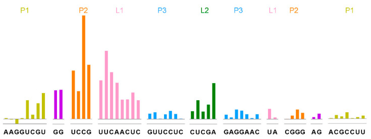
Figure 2.
Tetrahydrofolic acid recognizes the THF-II riboswitch. SHAPE analysis showed that tetrahydrofolic acid specifically recognizes the THF-II riboswitch Loop regions. The colored bars represent reduced SHAPE reactivity upon ligand binding. Residues are indicated on the X-axis. The coloring of the THF-II riboswitch with SHAPE signal is consistent with the secondary structure in Figure 1. The P1, P2 and P3 stems of the RNA are labeled in dark khaki, orange and blue, respectively. The GGAG-connected P1 and P2 are shown in purple. The Loop1 and Loop2 residues are shown in pink and green.

