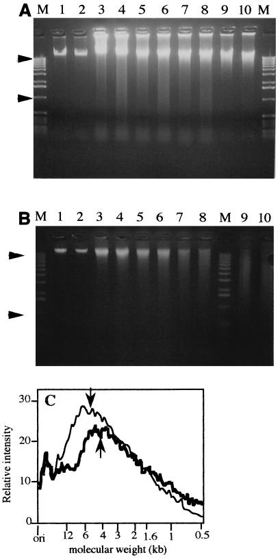
FIG. 1.
Chromosomal DNA extracted from UV-C-irradiated spores of strain WN356 electrophoresed through a 0.8% neutral agarose gel (A) and a 0.8% alkaline agarose gel (B). Spores were irradiated with 0 (lanes 1 and 2), 2 (lanes 3 and 4), 4 (lanes 5 and 6), 8 (lanes 7 and 8), or 16 (lanes 9 and 10) kJ of UV-C radiation per m2, and isolated DNA was treated with Endo V before electrophoresis (lanes 2, 4, 6, 8, and 10). Lanes M contained molecular weight markers (1-kb ladder; the arrowheads indicate the positions of 12- and 1-kb markers). (C) Densitometric scan of lanes 9 (thin line) and 10 (thick line) from the alkaline agarose gel shown in panel B containing DNA extracted from spores irradiated with 16 kJ of UV-C radiation per m2 before (lane 9) and after (lane 10) digestion with Endo V. The average single-strand fragment lengths were 4.7 kb before Endo V digestion (down arrow) and 4.2 kb after Endo V digestion (up arrow). ori, origin of gel.

