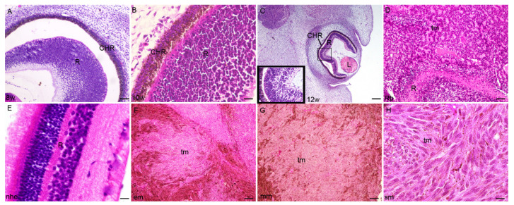
Figure 1.
Section through the eye of an 8th-week (8 w) (A), 10th-week (10 w) (B), and 12th week (12 w) (C) human embryo, retinoblastoma (Rb) (D), normal human eye (nhe) (E), epitheloid melanoma (em) (F), mixoid melanoma (mm) (G) and spindle melanoma (sm) (H); R—retina, CHR—choroidea, tm—tumor tissue, L—lens. Hematoxylin and Eosin staining, Scale bar for (A,D) = 50 μm, (B) = 25 μm, (E) = 10 μm, (C,F–H) = 100 μm.

