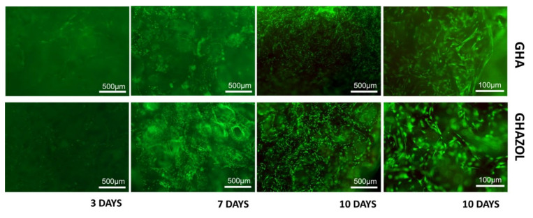
Figure 4.
Viability of materials. Live and dead cell staining was conducted on differentiated hMSC in the 3D fracture models treated with GHA and GHAZOL after 3, 7 and 10 days of culture. Viable cells stain green while dead cells stain red: images show that cells grew regularly into both 3D porous scaffolds as evident by the green fluorescent dye in absence of the red fluorescence dye (4× magnification: bar = 500 μm; 10× magnification: bar = 100 μm).

