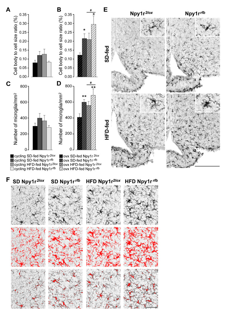
Figure 5.

Effect of HFD and Npy1r gene deletion on microglial activation and microglia number. The cell body to cell size ratio (cb/c) of microglia (A,B) and the number of microglia/mm2 (C,D). HFD significantly increased cb/c (B) and microglia number (D) in the ARC of ovx females. Conditional inactivation of Npy1r gene increased cb/c (B) and microglia number (D) in the ARC of SD- and HFD-fed ovx mice. Data are the mean ± SEM; n = 4–6 from 2 litters. # p < 0.05 versus SD-fed ovx females. * p < 0.05 and ** p < 0.01 versus Npy1r2lox ovx females on the same diet regimen. (E) Representative images of the ARC of ovx females expressing ionized calcium-binding adaptor protein-1 (IBA-1) immunoreactivity (scale bar: 150 μm). (F) Image analysis used to quantify the morphological characteristics of microglia in Iba-1 staining in ovx females. Upper panels: the unprocessed pictures. Middle panels: pixels darker than the background are traced (red) to determine the total cell size of microglia. Lower panels: pixel-clusters that are above an applied staining threshold and size-filter are traced (red) to determine the total cell body size of all microglia, as well as the number of microglia.
