Figure 1.
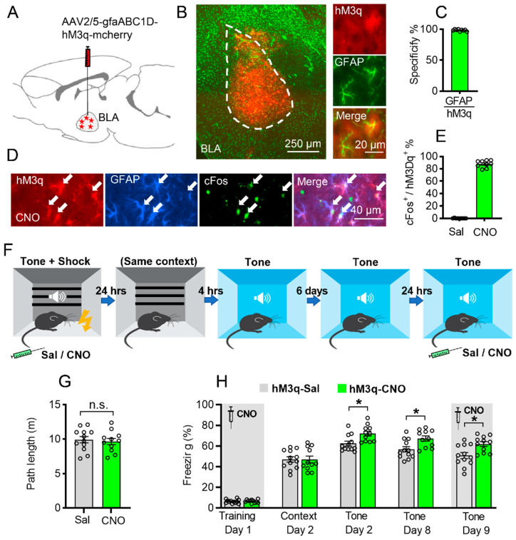
Astrocytic Gq activation in the BLA promotes auditorily cued fear memory formation. (A) Schematic picture showing AAV-gfaABC1D-hM3q-mcherry injection in the BLA bilaterally. (B) hM3q virus expression in the BLA (left panel); red is hM3q, green is GFAP staining, scale bar 250 µm. Magnified view of hM3q co-expressed with GFAP (right panel); scale bar 20 µm. (C) Cells with hM3q expression are highly co-labeled with GFAP (98.33 ± 0.5%, n = 3, 9 slices). (D,E) CNO administration to mice expressing hM3q (red) in BLA astrocytes resulted in a significant increase in c-Fos expression (green) in these astrocytes (white arrow), compared to saline-injected controls (p < 0.0001, n = 3 per groups, 9 slices per group; blue is GFAP staining, scale bar 40μm). (F) Schematic diagram of fear memory training and contextual/auditorily cued fear memory retrieval. (G) Path length of exploration in the conditioning cage after CNO or saline injection (p > 0.05, unpaired Student’s t-test, n.s. stand for no significance). (H) Chemogenetic activation of BLA astrocytes increased auditorily cued fear memory on day 2 (p = 0.0367), day 8 (p = 0.0424), but not contextual fear memory (p > 0.05) compared to saline group. On day 9, CNO injection before auditorily cued memory test still showed significant enhancement of fear memory compared to saline group (p = 0.0361) (Saline group n = 12, CNO group n = 11, * stands for p < 0.05, two-way ANOVA followed by Bonferroni’s post hoc test, main effect of Sal/CNO: F (1, 21) = 7.530, p = 0.0122). Data are presented as mean ± SEM.

