Figure 3.
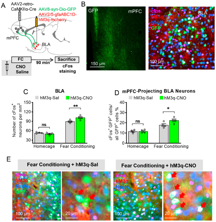
Astrocytic Gq modulation induces a projection-specific enhancement of BLA–mPFC neurons during fear learning. (A) Schematic diagram of targeting mPFC-projecting BLA neurons: AAV2-retro-CamkII-Cre was injected into the mPFC, and AAV8-syn-Dio–GFP together with AAV2/5-gfaABC1D-hM3q-mcherry was injected into the BLA. (B) Left: GFP-positive axons of BLA projection neurons are clearly visible in the mPFC. Right: mPFC-projecting BLA neurons (green) with c-Fos (pink) and NeuN (blue) staining, and hM3q (red) in BLA astrocytes. (C) c-Fos level in BLA neurons of four-group mice expressing GFP in mPFC-projecting BLA neurons and hM3q in their BLA astrocytes (n = 4 of each group, 5 slices for each mouse, two-way ANOVA with Bonferroni post hoc comparison test, main effect of Sal/CNO: F (1, 6) = 6.423, p = 0.0444, ‘homecage (HC) + hM3q-Sal’ versus ‘HC + hM3q-CNO’: p > 0.9999, ‘fear conditioning (FC) + hM3q-Sal’ versus ‘FC + hM3q-CNO’: p = 0.0055, ** stands for p < 0.01, ns stands for no significance). (D) Fear-conditioned mice injected with CNO showed increase in the percentage of mPFC-projecting BLA neurons that express c-Fos compared with FC mice injected with saline (n = 4 of each group, two-way ANOVA with Bonferroni post hoc comparison test, main effect of Sal/CNO: F (1, 6) = 7.424, p = 0.0344, ‘HC + hM3q-Sal’ versus ‘HC + hM3q-CNO’: p > 0.9999, ‘FC + hM3q-Sal’ versus ‘FC + hM3q-CNO’: p = 0.032, * stands for p < 0.05, ns stands for no significance). (E) Representative images of hM3q (red), c-Fos (pink), mPFC-projecting BLA neurons (green), and NeuN (Blue) in the BLA; red arrow shows the mPFC-projecting BLA neurons with c-Fos expression. All data are presented as mean ± SEM.

