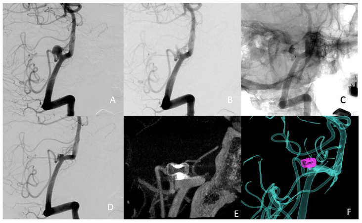
Figure 5.
(A) preoperative treatment angle; (B) subtracted postoperative image; (C) nonsubtracted postoperative image, showing the stagnation of the contrast agent in the dome of the aneurysm, suggesting successful occlusion (D) 3-month follow-up treatment angle (E) 3-month follow-up 3D angio-CT (F) 3-month follow-up fusion imaging.

