Figure 1.
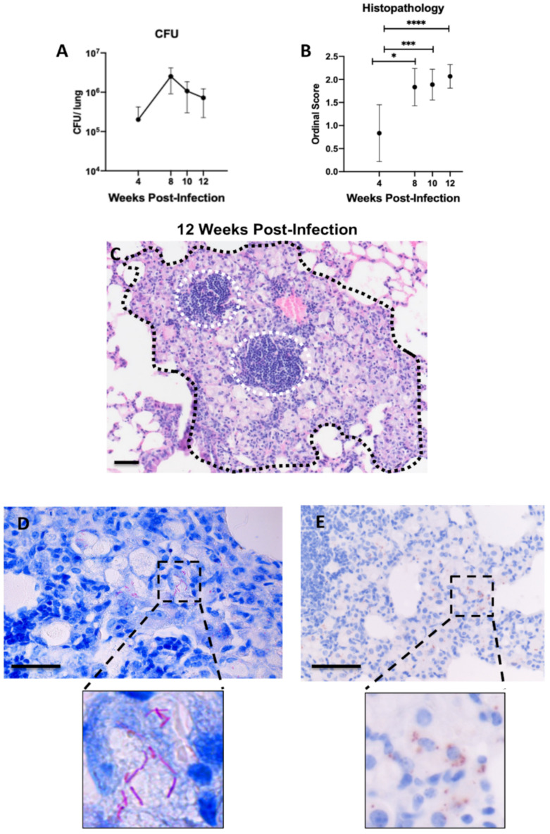
Establishment of chronic pulmonary M. avium infection with intranasal inoculation 4–12 weeks post-infection (wpi) in B6.Sst1S mice. (A) CFU/lung following intranasal (IN) 106 inoculation; (B) histopathology scores of 106 CFU IN M. avium inoculated mice at 4, 8, 10, and 12 wpi; (C) representative histological micrograph of granulomatous lesion (black hash) with intralesional lymphoid aggregates (white hashed circles) caused by M. avium at 12 wpi assessed by hematoxylin and eosin (H&E) and visualized using brightfield microscopy; (D) detection of M. avium Acid-fast bacilli using Ziehl-Neelsen stain and visualized using brightfield microscopy; (E) diaminobenzidine (DAB) immunohistochemistry detection of Mycobacterial antigen visualized using brightfield microscopy. High magnification insets are located below (D,E) to highlight distinct differences in these modalities utilized to visualize mycobacterium bacilli and antigen respectively. For (A), each data point represents average CFU/lung; n = 6 (4 wpi), n = 2 (8 wpi), n = 3 (10, 12 wpi). For (B), each data point represents the mean from a single tissue section; n = 18 (4 wpi), n = 6 (8 wpi), n = 9 (10 wpi), n = 15 (12 wpi). Data are expressed as means ± SD. * p < 0.05, *** p < 0.0005, and **** p < 0.00005. Original magnification, 100× (C) and 200× (D,E); scale bars = 50 μm.

