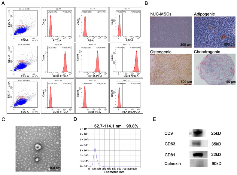
Figure 1.
Identification and characterization of hUC-MSCs and secreted EVs. (A) Identification of surface marker proteins of hUC-MSCs by flow cytometry. (B) The differentiation ability of hUC-MSCs was evaluated by cell staining. (C) The morphological characteristics of the EVs were observed by TEM. (D) The particle size distribution range of the EVs was analyzed by NTA. (E) The surface markers of EVs were analyzed by Western blot.

