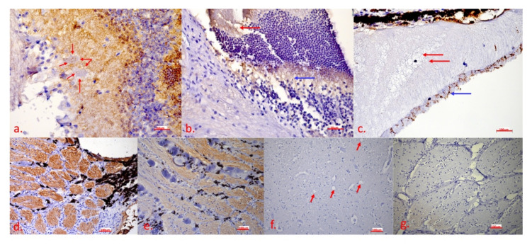
Figure 6.
IHC detection of caspase 3 shows the presence of apoptosis in the eye bulb and the brain: (a) retina—strong presence of the brown chromogen in the inner plexiform layer, presence of the brown chromogen in the ganglion cells (red arrows); (b) optic disc—presence of the brown chromogen in the cone and rod layer (red arrow) and the outer plexiform layer (blue arrow); (c) subchoroidal layer/retina—oedema (red arrow) between the pigment epithelium and the cone and rod layer, presence of the brown chromogen in the cone and rod layer (blue arrow); (d) optic disc—strong presence of the brown chromogen in nerve fibres; (e) optic disc—calcifications between nerve fibres; (f) brain—no apoptosis signalling, vacuoles are marked with the red arrow; (g) optic disc—negative control. In Figure 6a,f, the scale bar corresponds to 50 μm (40× magnification), in Figure 6b,c,d,e,g, the scale bar corresponds to 100 μm (20× magnification). IHC to detect caspase 3, DAB chromogen, counterstain with Mayer’s haematoxylin.

