Figure 1.

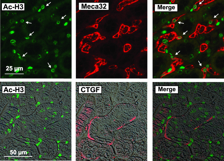
Detection of CTGF and acetylated histone H3 in mouse kidney sections. Sections of mouse kidneys were double‐stained for acetylated histone H3 (Ac‐H3) and the endothelial cell marker Meca32 (upper panels) or Ac‐H3 and CTGF (lower panels). Images taken with polarization filters were used as background in the lower panels to show the localization of CTGF staining. Data are representative of eight individual kidneys. Arrows indicate cells positive for Ac‐H3 and Meca32.
