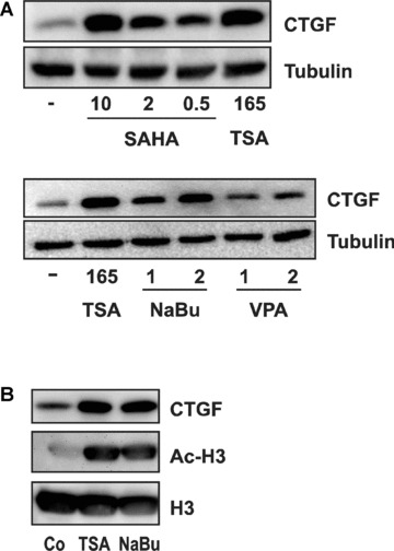
Figure 3.

Induction of CTGF by chemically different HDIs. (A) glEND.2 cells were stimulated for 24 hrs with different HDAC inhibitors: TSA (165 nM), SAHA (0.5, 2 and 10 μM), sodium butyrate (NaBu, 1 and 2 mM) and valproic acid (VPA, 1 and 2 mM). Cellular extracts were separated by SDS PAGE and CTGF protein was detected by Western blotting. (B) glEND.2 cells were stimulated with TSA (165 nM) or sodium butyrate (NaBu, 2 mM) for 6 hrs. Histone H3 and acetylated histone H3 (Ac‐H3) were detected in nuclear extracts, whereas CTGF was detected in the cytosolic fraction.
