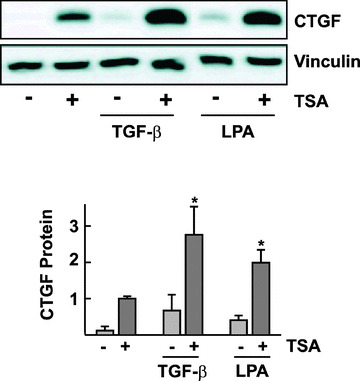
Figure 4.

Facilitation of TGF‐β‐ and LPA‐mediated CTGF protein expression by TSA. glEND.2 cells were pre‐incubated for 20 hrs with TSA (330 nM) and then stimulated with TGF‐β (TGF‐β, 5 ng/ml, 5 hrs) or LPA (LPA, 10 μM, 2 hrs). CTGF protein from cellular homogenates was analyzed by Western blotting. The graph summarizes the results of n= 4 experiments; *P < 0.05 ANOVA with Tukey’s post‐test, TSA plus TGF‐beta or TSA plus LPA compared with any stimulus alone,
