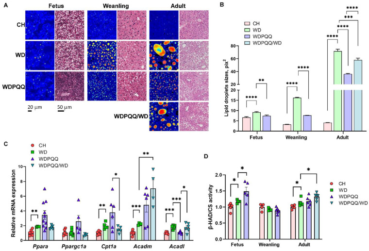
Figure 2.
Hepatic steatosis in WD-fed offspring is improved by PQQ. (A) FLIM images and H&E staining of liver cryosections in fetuses (E18.5), weanlings (3 wks of age), and adults (20–24 wks of age). Scale bars: 20 μm for FLIM and 50 μm for H&E stains. Images acquired at 400× final magnification for FLIM and 40× for H&E. (B) Lipid droplet areas were quantified using ImageJ. Data are means ± SEM. ** p < 0.01, *** p < 0.001, **** p < 0.0001. (C) qPCR of genes involved in fatty acid oxidation in adult liver tissue. 18S was used as a reference gene. Data are means ± SEM. ** p < 0.01, *** p < 0.001. (D) Activity of β-HAD in liver homogenates. Citrate synthase (CS) activity was used for normalization. Data are means ± SEM. * p < 0.05 compared with WD.

