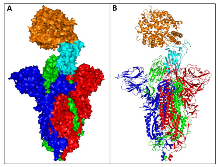
Figure 1.
The crystal structure of SARS-CoV-2 S protein complexed with ACE2 receptor retrieved from the Protein Data Bank (PDB), PDB entry 7DF4. The structure was visualised by PyMOL (The PyMOL Molecular Graphics System, Version 1.7.4 Schrödinger, LLC.). The complex is displayed as (A) surface and (B) loops. The S protein assembles into trimers (coloured red, blue, and green) on the virion surface to form a distinctive “corona”. The RBD domain of the S protein (cyan) binds to the human ACE2 receptor (orange) to promote attachment and fusion.

