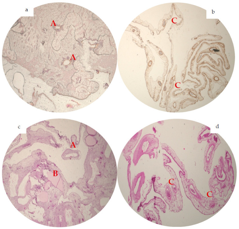
Figure 2.
Representative immunohistochemical analysis of angiogenesis (2× magnification, n = 48). In alphaSMA staining, BSM group (a) showed solitary big-diameter vessels (A) and some small-diameter vessels. The alphaSMA staining of BSM+ (b) presented numerously small-diameter vessels, attaching prominently to the BSM+ (C). On the H&E staining, BSM (c) displays bigger diameter vessels (A) and almost no small-diameter vessels with a vessel grown through the BSM (B), in the H&E slices of BSM+ (d) a great number of a small- and medium-diameter vessels could be observed (C).

