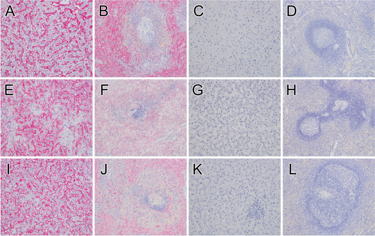
Fig 2. Immunohistochemistry of liver and spleen tissue in MARV-infected macaques.
MARV antigen (red) IHC positivity in sinusoidal lining cells and Kupffer cells of the liver for the positive control (Control 1) (A), group day -5 vaccinated fatal subject (Fatal 1) euthanized 9 DPI (E), group day -3 vaccinated fatal subject (Fatal 4) euthanized 7 DPI (I). IHC positive mononuclear cells within the red and white pulp of the spleen for the positive control (Control 1) (B), group day -5 vaccinated fatal subject (Fatal 1) euthanized 9 DPI (F), group day -3 vaccinated fatal subject (Fatal 4) euthanized 7 DPI (J). No appreciable immunolabeling of the liver or spleen for macaques that survived from group day -7 (Survivor 1) (C, D), group day -5 (Survivor 7) (E), or group day -3 (Survivor 10) (I). All representative liver sections were taken at 20x magnification, and all spleen sections were taken at 10x magnification. IHC, immunohistochemistry; MARV, Marburg virus; DPI, days post infection.

