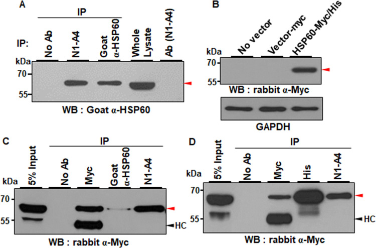
Fig 3. N1-A4 recognizes HSP60.
(A) Huh7 cell lysates were immunoprecipitated with N1-A4 or goat anti-HSP60 antibodies (α-HSP60), and the immunoprecipitates were detected with α-HSP60 in Western blot analysis. Red arrowhead indicates HSP60. (B) HSP60-Myc/His vector was overexpressed in 293FT cells, and Myc-tagged HSP60 protein was detected with α-Myc. β-actin expression was the loading control. (C, D) HEK293FT cells were transfected with HSP60-Myc/His expression vector. Cell lysates were immunoprecipitated with α-Myc, α-HSP60, and N1-A4 (C), or α-Myc, α-His, and N1-A4 (D). The immunoprecipitates were detected by Western blot analysis with α-Myc. Red arrowhead indicates the HSP60 proteins.

