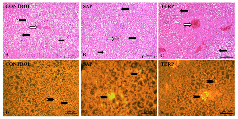
Figure 4.
Representative pictures of rat liver stained with hematoxylin and eosin ((A–C) upper panel) and PMP70 immunohistochemical staining for the 70 kDa peroxisomal membrane protein marker PMP70 ((D–F) yellow signals in lower panel; solid arrowhead = positive signals detecting the presence of the peroxisomes in controls and experimental groups). Note the degree of steatosis—different amounts and sizes of the fat droplets in the hepatocytes (solid arrows = hepatocytes; open arrows = central vein) in pictures stained with hematoxylin and eosin. Mag. 200×. Scale bar 100 µm (A–F).

