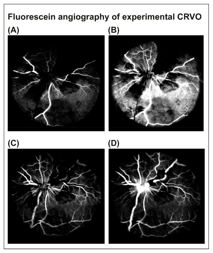
Figure 2.
Fluorescein angiography was performed 3 days after CRVO to confirm that the occlusion was successfully induced. (A–C) Early phases of fluorescein angiography of experimental CRVO at 15–24 s. Angiography revealed tortuous retinal veins, delayed venous filling and retinal capillary non-perfusion following CRVO. (D) Angiography at 1 min and 26 s. Venous dilation, retinal capillary non-perfusion and leakage of fluorescein were observed.

